Cutaneous Squamous Cell Carcinoma
Squamous Cell Carcinoma (SCC) is a malignant epithelial tumor of the skin.
- It grows both in width and in depth, although it often develops as an exophytic tumor, meaning that it protrudes above the skin.
- It originates from the squamous cells of the epidermis and the skin appendages.
- It may affect both the skin and the mucous membranes.
- It may develop de novo or on a background of actinic keratosis.
- It usually appears as a superficial lesion with a firm texture, resembling a wart or a scaly plaque.
- It may ulcerate and is often accompanied by itching.
- It may bleed very frequently.
- It may become infected and resemble a wound that discharges fluid or pus.
- It is usually painless, but when it grows significantly, it may become painful.
- It may metastasize either locally or to distant organs.
Because of the risk of metastasis and the generally aggressive behavior of squamous cell carcinomas, their treatment must be radical.
Cutaneous Squamous Cell Carcinoma – When Does the Patient Visit the Doctor?
Patients with squamous cell carcinoma usually delay visiting the doctor. The reason is that, in most cases, the tumor grows slowly and is usually painless.
Patients visit the doctor when:
- the tumor begins to grow rapidly in size (this usually happens suddenly within the last 2 to 3 months, while initially the tumor remained small for a long period)
- the tumor begins to bleed
- the tumor discharges pus
- the tumor develops an unpleasant odor
- the tumor becomes painful
Cutaneous Squamous Cell Carcinoma – Incidence
Squamous Cell Carcinoma is the second most common type of skin cancer after Basal Cell Carcinoma (BCC). According to international statistics, approximately 450,000 new cases are diagnosed every year.
We should keep in mind that among skin cancers:
- 10% are Squamous Cell Carcinomas
- 85% are Basal Cell Carcinomas
- 3 to 4% are melanomas, and
- 1 to 2% are unclassified tumors.
Cutaneous Squamous Cell Carcinoma usually appears during the seventh decade of life, while it is extremely rare before the age of 50. Men are generally affected twice as often as women.
Cutaneous Squamous Cell Carcinoma – Causes
Cutaneous Squamous Cell Carcinoma is named after the squamous cells from which it originates, meaning cells located in the spinous layer of the epidermis.
It is a tumor that develops in the skin and is directly associated with exposure to solar radiation. The more someone has been exposed to the sun, the greater their risk of developing Squamous Cell Carcinoma, as well as Basal Cell Carcinoma, of the skin.
Apart from sun exposure, other causes of Squamous Cell Carcinoma include:
- exposure to artificial tanning (solarium)
- old injuries
- old burns (Marjolin ulcer)
- scars and skin ulcers
- chronic infections
- immunodeficiency, such as in patients with HIV.
Cutaneous Squamous Cell Carcinoma – Who Does It Affect?
There is one simple rule:
The more you are exposed to the sun, the greater your risk of developing skin cancer. If there is also a genetic predisposition, the risk is even higher.
Therefore, everyone who is exposed to the sun for many hours, such as laborers, construction workers, farmers, etc., as well as individuals who spend many hours sunbathing, are at high risk of developing Squamous Cell Carcinoma.
It usually affects elderly individuals after the seventh decade of life and is more common in people with blond hair and blue or green eyes.
If someone develops a Basal Cell Carcinoma on the skin, they have a significantly greater chance of also developing a Squamous Cell Carcinoma.
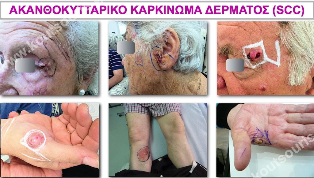
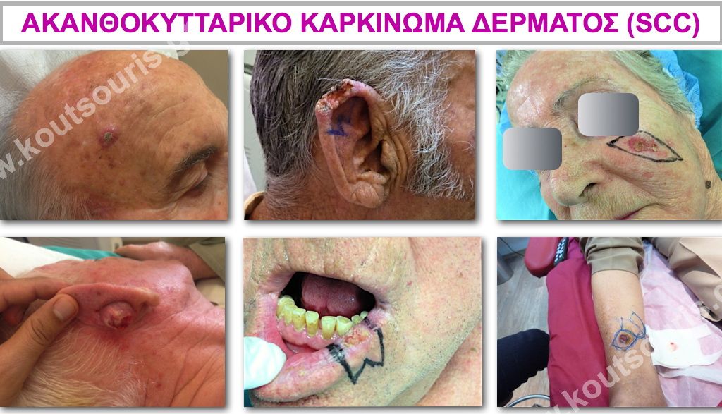
Cutaneous Squamous Cell Carcinoma – Where Does It Appear?
It appears on areas of the skin that are exposed to the sun. It may also affect the mucous membranes of both the mouth and the genital organs.
The most common locations are:
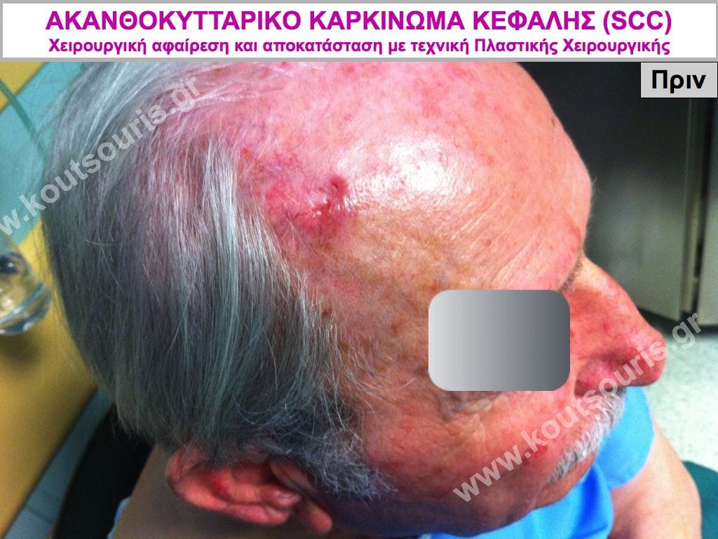
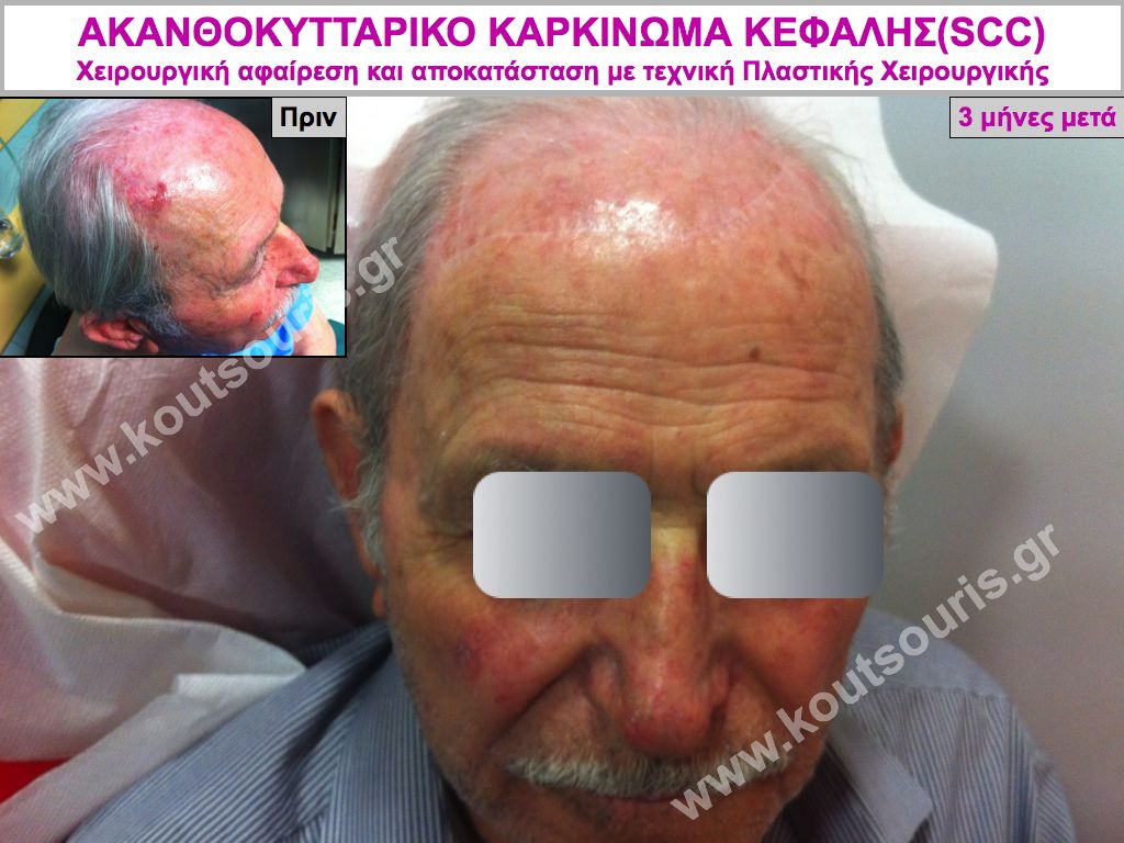
- the bald area of the scalp
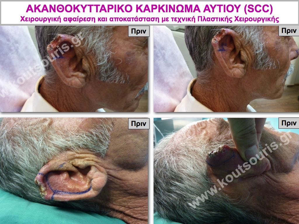
- the outer ear, usually the upper part
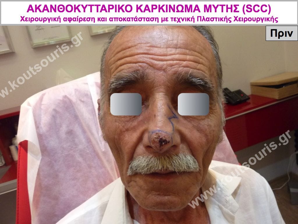
- the bridge of the nose
- the lower lip
- the cheek
- the lower eyelids
- the forehead
- the shoulders
- the hands
- the front surface of the shin
- old ulcers, wounds or burns (Marjolin ulcer)
- the mucous membranes of the genital organs
- the oral mucosa.
It Often Begins From Precancerous Lesions
Squamous Cell Carcinoma may begin from precancerous lesions or atypical cancerous growths.
Such precancerous skin lesions include:
- Actinic or solar keratoses.
These are rough, slightly raised or blister-like lesions that usually develop on areas of the skin exposed to the sun.
Today, we know that 45 to 65% of squamous cell carcinomas develop on a background of actinic keratoses.
Therefore, when these lesions appear on the skin, they should be treated immediately; otherwise, they may develop into aggressive skin cancer.
- Actinic cheilitis.
This is a type of actinic keratosis. It usually appears on the lower lip rather than the upper lip because of direct exposure to solar radiation. Clinically, it is characterized by dryness and cracking of the lip mucosa.
- Leukoplakia
These are white patches on the oral mucosa, tongue or gums. It is a condition associated with excessive alcohol consumption, smoking or frequent trauma inside the mouth and may lead to squamous cell carcinoma.
- Bowen’s disease.
It appears as a reddish-brown scaly plaque resembling eczema. It is a superficial lesion that nevertheless has invasive tendencies and is considered a form of early-stage, non-invasive squamous cell carcinoma.
Bowen’s disease is associated with sun exposure, as well as substances containing arsenic or other carcinogenic chemical agents. Infection with the human papillomavirus (HPV) following sexual contact may also lead to the development of Bowen’s disease on the genital organs.
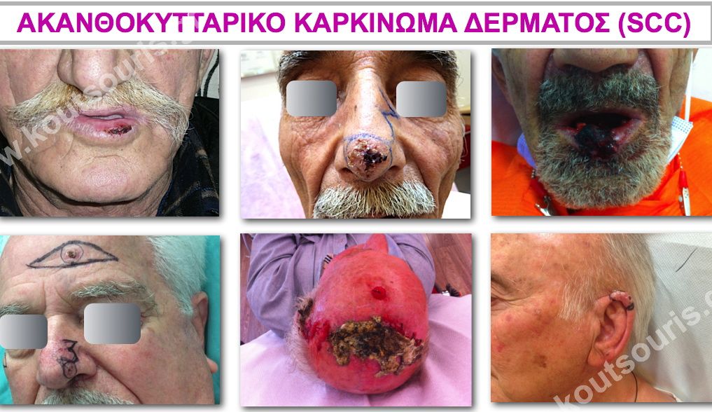
Cutaneous Squamous Cell Carcinoma – Appearance / Progression
Squamous Cell Carcinoma most often appears as a dirty-looking wound on the skin with raised edges. It often bleeds if accidentally injured and may be covered by a crust, beneath which clear or yellowish fluid is discharged.
It is a wound that does not heal with antibiotic ointments, grows by eroding the surrounding tissues and often deforms the affected area.
It usually takes a long time, months or even years, to grow.
Initially, it is very small and grows slowly. However, once it reaches a significant size, the tumor may double or triple in size very rapidly, within 2 to 3 months. This is usually when the patient becomes concerned and visits the doctor.
The appearance of a wound that “closes and then opens again”, despite careful local treatment, should make us suspect that something serious may be occurring. Furthermore, as mentioned above, if it bleeds following minor trauma, we should again be concerned and seek immediate treatment.
Cutaneous Squamous Cell Carcinoma – Treatment Options
Squamous Cell Carcinoma is an aggressive form of skin cancer. Apart from local tissue erosion and destruction of the skin, the main concern is that it may also metastasize. We must not forget this and should therefore be extremely careful in its treatment.
However, if Squamous Cell Carcinoma is removed promptly, it can be cured. The essential condition is radical removal of the tumor so that the remaining tissues are 100% healthy.
The treatments recommended for Squamous Cell Carcinoma are:
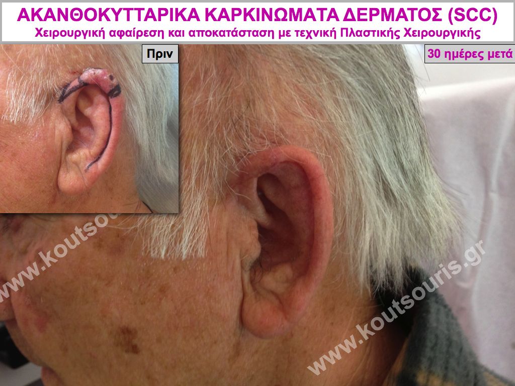
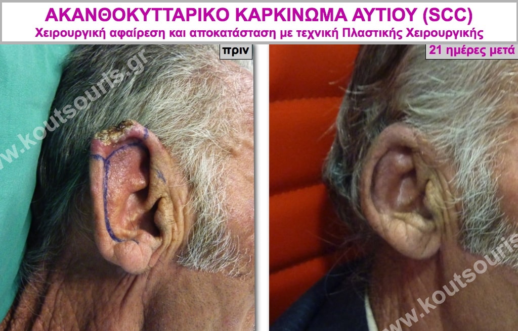
- surgical excision of the tumor with biopsy and plastic reconstruction
- electrocautery
- cryotherapy
- photodynamic therapy (PDT)
- topical application of pharmaceutical preparations
- Laser cauterization
- radiation therapy.
Cutaneous Squamous Cell Carcinoma – Advantages of Surgery
When surgically removing a skin lesion or lump using a scalpel, we completely remove the tumor and immediately send it for frozen-section biopsy. Once the pathologist confirms during surgery that the cancer has been completely removed, we can safely proceed with closure of the defect using plastic reconstruction.
Surgical removal of a tumor and its immediate pathological examination with frozen-section biopsy offers the advantage that:
- through frozen-section biopsy, we can immediately determine whether the lesion is cancerous and identify the type of cancer, and
- we can be certain that the cancer has been completely removed and therefore speak of a cure immediately after the procedure.
However, in patients who cannot undergo surgery because they suffer from serious accompanying health conditions, such as cardiac or hematological problems, immunosuppression, very advanced age, etc., we proceed with radiation therapy or other non-surgical treatments.
Squamous Cell Carcinoma – Disadvantages of Non-Surgical Treatments
When treating skin cancer using ointments, cryotherapy or cauterization, meaning by destroying the tumor, there is the problem that WE DO NOT HAVE IDENTIFICATION OF THE LESION THROUGH BIOPSY.
The principle of non-surgical treatments is that we destroy the affected area as extensively as possible in order to eliminate the cancerous foci. However, with non-surgical treatments, we can never be certain that we have destroyed 100% of all cancer cells, since we do not have the removed tumor available for examination.
Therefore, if the cancer has not been completely destroyed, it will recur within a relatively short period of time and, in most cases, will be more aggressive.
This is the problem with non-surgical treatments and the reason why we prefer surgical treatment.
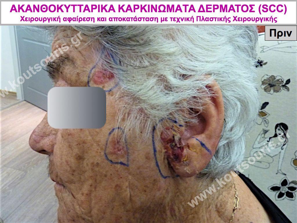
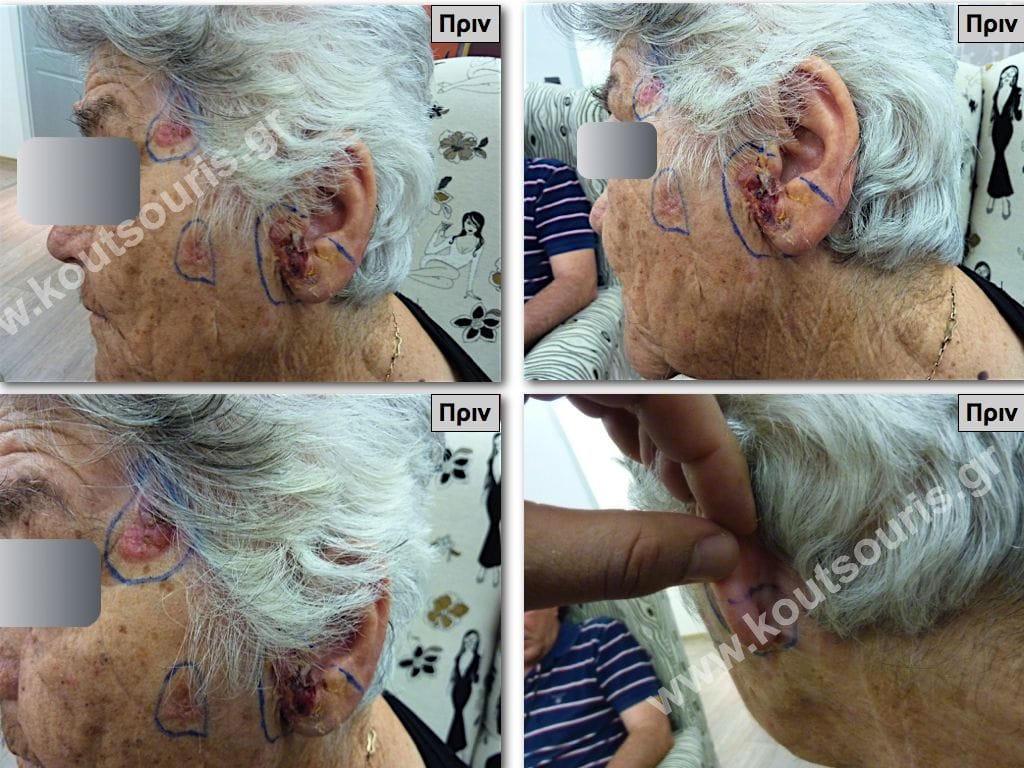
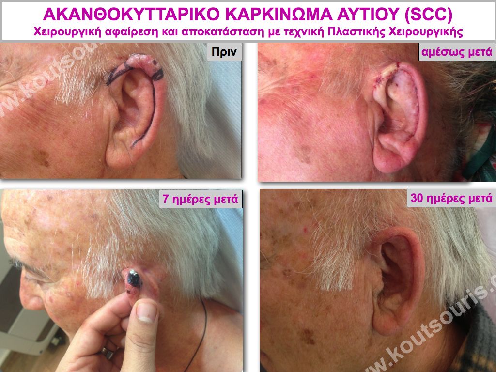
Cutaneous Squamous Cell Carcinoma – Surgical Removal / Plastic Reconstruction
We recommend treating skin cancer through surgical removal of the tumor followed by plastic reconstruction of the defect.
Small cancers are usually removed under local anesthesia in the medical office.
Medium-sized and large tumors are treated in an organized surgical environment, and frozen-section biopsy is always performed during the procedure.
Cutaneous Squamous Cell Carcinoma – How Is Surgery Performed?
It is an almost painless procedure. Most of the time, it is performed under local anesthesia while the patient remains awake. In very rare cases, it may be performed using a combination of local anesthesia and sedation, or general anesthesia may be required.
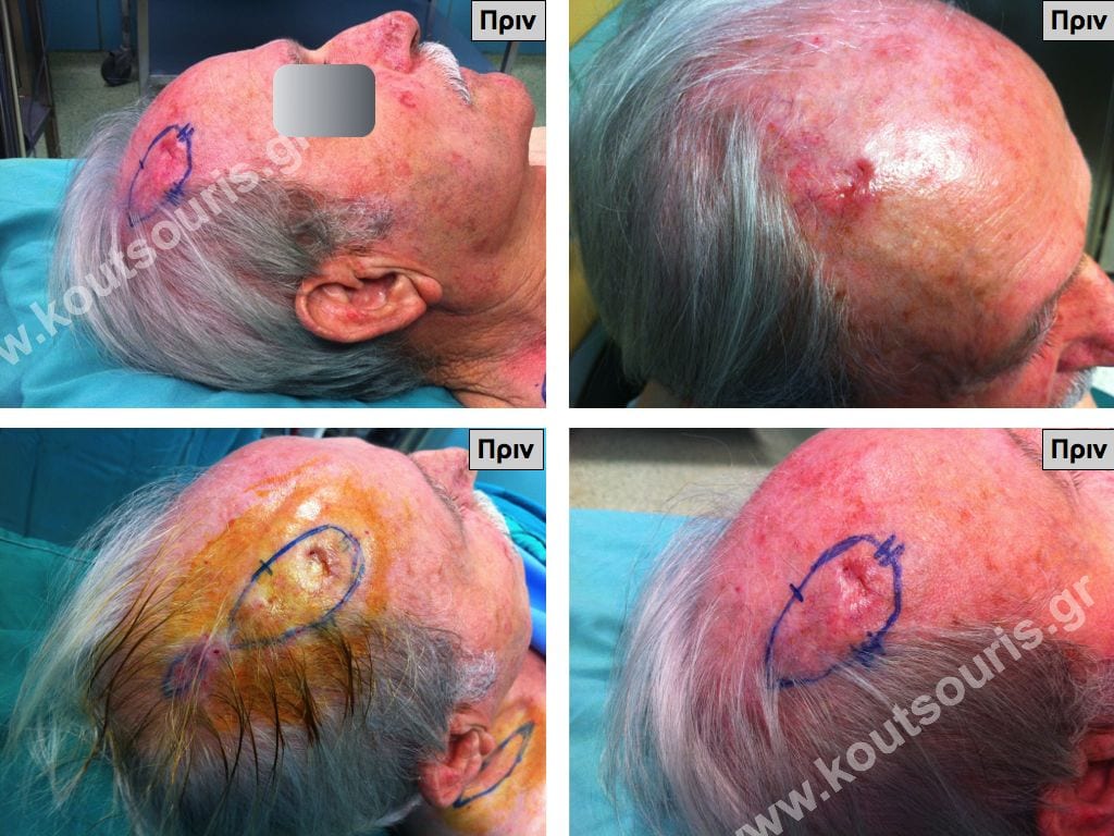
The main objective of surgery is radical excision of the tumor. For this reason, we always design the excision leaving 3 to 5 millimeters of healthy tissue around the lesion.
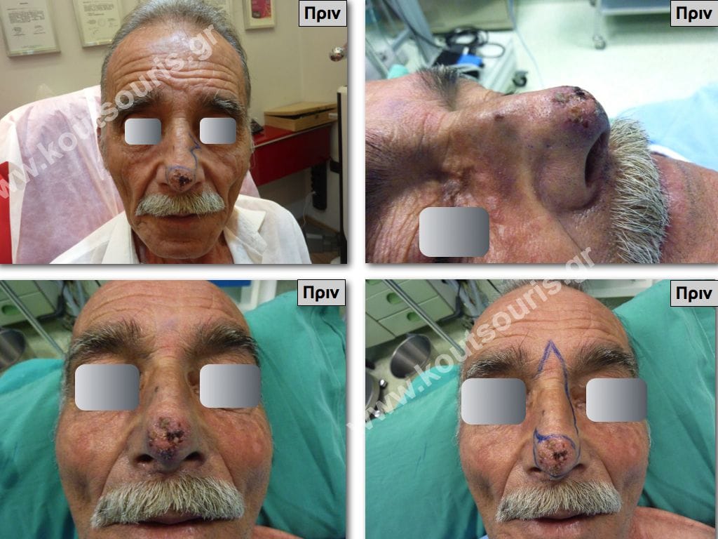
We always map the cancer using the clock-face marker technique at the 3, 6, 9 and 12 o’clock positions. In this way, the pathologist can identify the areas where the tumor is developing within the biopsy specimen.
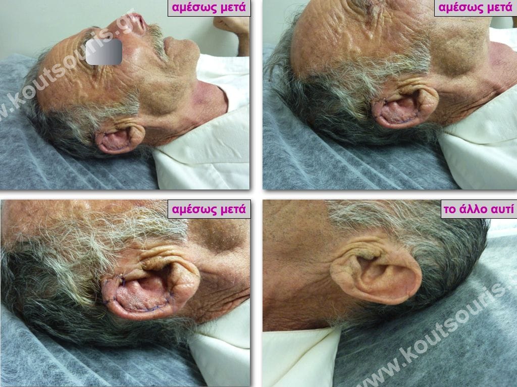
After mapping the lesion, we design the plan for plastic reconstruction.
The area is then anesthetized and the tumor is widely excised. The specimen is subsequently sent for frozen-section biopsy.
Usually, 15 to 20 minutes are sufficient to receive the frozen-section biopsy result. What we need to determine is whether the lesion is cancerous, the type of cancer and, most importantly, whether the lesion has been completely removed within healthy tissue margins.
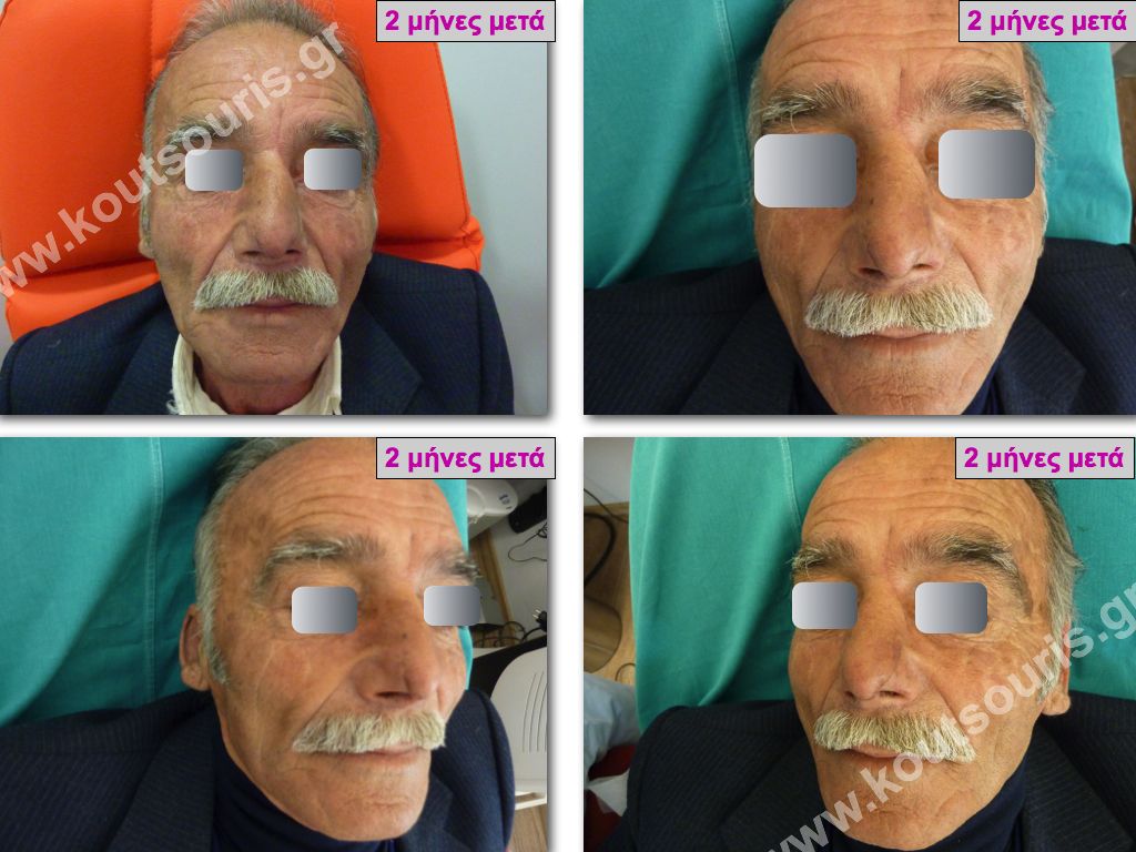

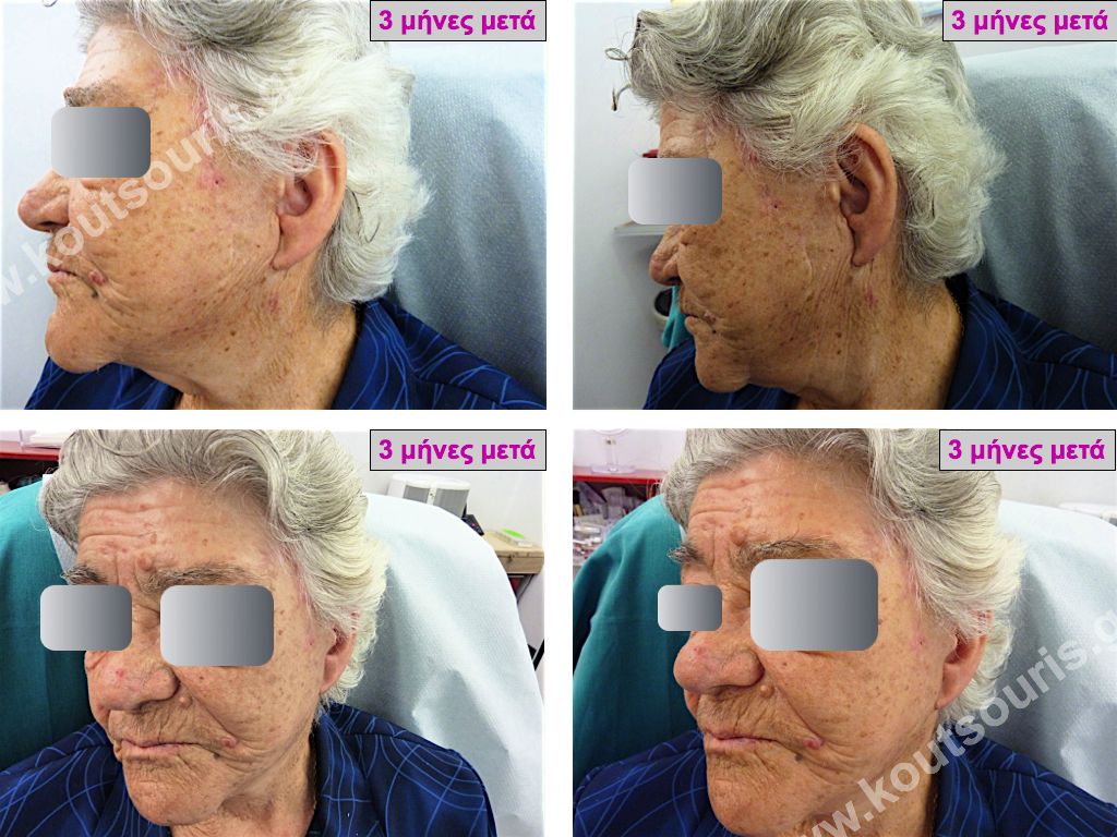
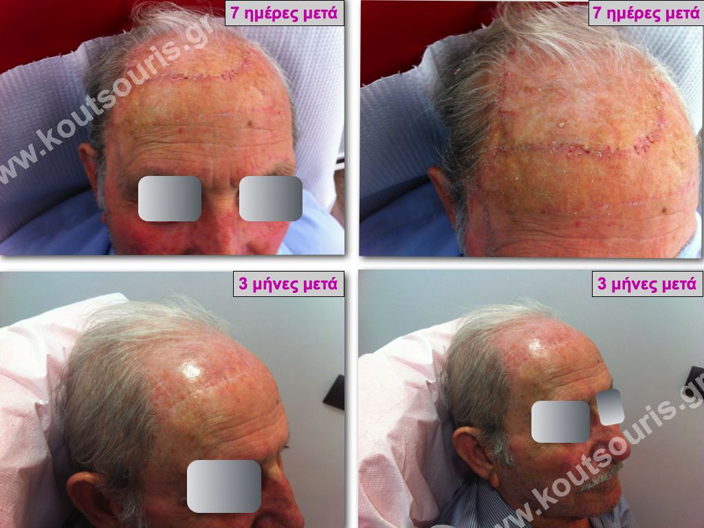
If the pathologist confirms that the lesion has been completely removed, we proceed with plastic reconstruction.
If the result indicates that cancerous tissue remains, we immediately proceed with additional surgical removal from the margins of the previous excision. We send another frozen-section biopsy and, once we are certain that no cancerous foci remain, we close the wound using plastic reconstruction.
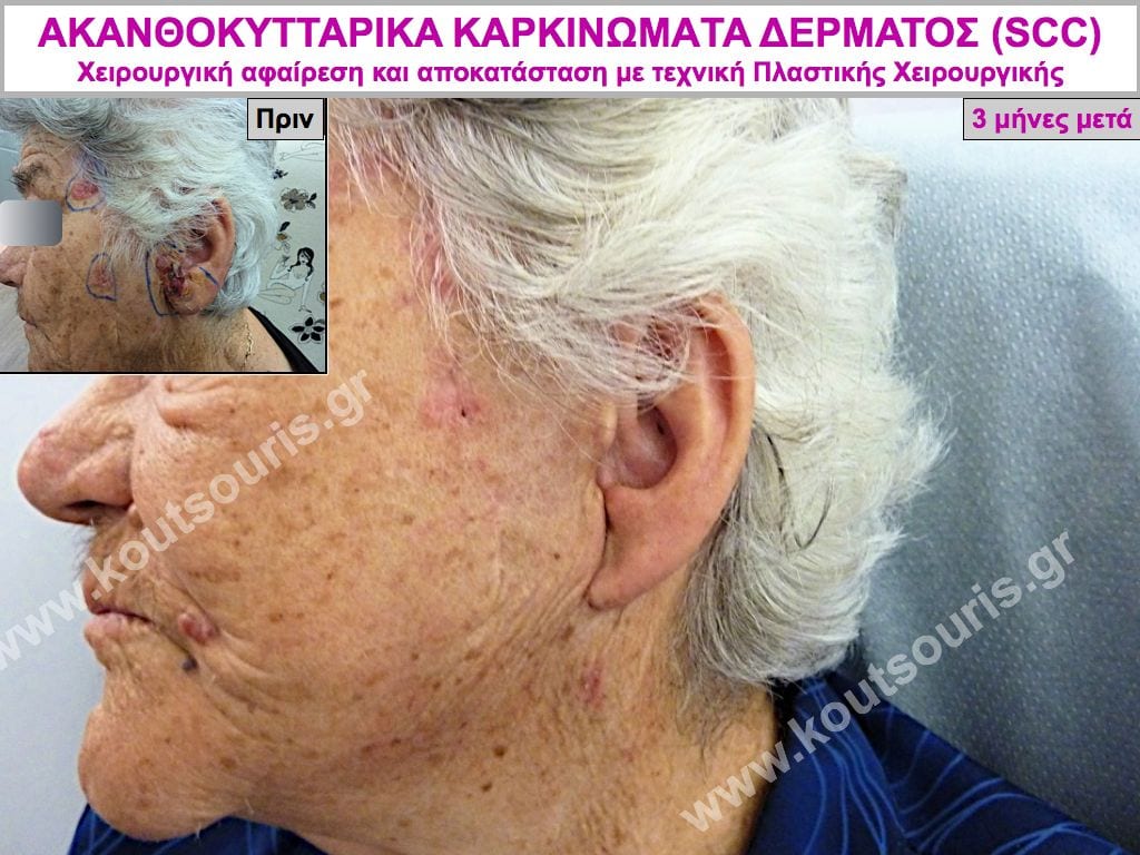
Using this surgical technique, we can achieve a high level of safety and speak of a cure in most cases of skin cancer.
After Surgery for Cutaneous Squamous Cell Carcinoma
The fundamental principle is that the skin cancer must have been removed within healthy tissue margins.
In general, no additional treatment with chemotherapy, radiation therapy or other accompanying treatments is required.
Only immediate examination for possible metastases and preventive periodic monitoring of the patient for at least one to two years are required.
Cutaneous Squamous Cell Carcinoma – Prognosis
Squamous Cell Carcinoma is a malignant disease of the skin. It remains localized in the epidermis for a long period but subsequently infiltrates and “eats away” the surrounding tissues, causing destruction and major deformities in the area where it appears.
Squamous Cell Carcinomas may metastasize in approximately 10-12% of cases, both to neighboring tissues and distant organs, and may threaten the patient’s life if left untreated. Today, we know that approximately 2,500 patients die every year in the United States.
If the tumor is removed promptly within healthy tissue margins, the specific lesion can be considered cured. However, we should keep in mind that when a patient develops Cutaneous Squamous Cell Carcinoma on sun-damaged skin, there is a high likelihood of developing a second cancer in a neighboring area or even in the same area, as well as another cancer elsewhere on the body.
This may occur even though the original tumor was completely removed surgically. The explanation is that the skin has sustained permanent and extensive sun damage, and independent cancers may develop in the surrounding areas. Recurrences usually appear within the first two years.
Squamous Cell Carcinomas located on the ears, nose and lips have the greatest likelihood of recurring, even after surgical removal.
All patients who have developed Squamous Cell Carcinoma should be examined periodically for possible new skin lesions. Prevention, early diagnosis and appropriate treatment are therefore extremely important for the prognosis of this type of cancer.
Cutaneous Squamous Cell Carcinoma – What Should We Keep in Mind?
Any “spot” or open wound that does not heal within three weeks despite treatment with topical antiseptics has a high likelihood of being skin cancer.
The patient must be examined by a specialized doctor and treated without any delay.
Early diagnosis and appropriate surgical treatment lead to a cure and help prevent serious complications.
BEFORE & AFTER CASES